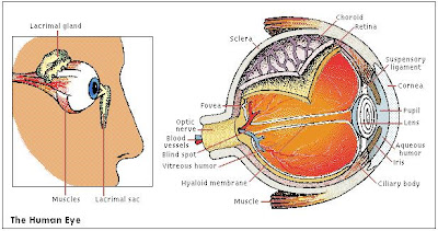
Posts

Brain

Image
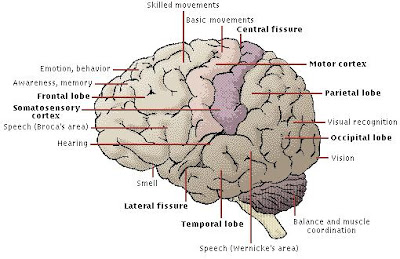
Brain, portion of the central nervous system contained within the skull. The brain is the control center for movement, sleep, hunger, thirst, and virtually every other vital activity necessary to survival. All human emotions —including love, hate, fear, anger, elation, and sadness—are controlled by the brain. It also receives and interprets the countless signals that are sent to it from other parts of the body and from the external environment. The brain makes us conscious, emotional, and intelligent. THE HUMAN BRAIN The human brain has three major structural components: the large dome-shaped  cerebrum (top), the smaller somewhat spherical cerebellum (lower right), and the brainstem (center). Prominent in the brainstem are the medulla oblongata (the egg-shaped enlargement at center) and the  thalamus (between the medulla and the cerebrum). The cerebrum is responsible for intelligence and reasoning. The cerebellum helps to maintain balance and posture. The medulla is involve

The Human Eye

Image
Structure of the Eye The amount of light entering the eye (right) is controlled by the pupil, which dilates and contracts accordingly. The cornea and lens, whose shape is adjusted by the ciliary body, focus the light on the retina, where receptors convert it into nerve signals that pass to the brain. A mesh of blood vessels, the choroid, supplies the retina with oxygen and sugar. Lacrimal glands (left) secrete tears that wash foreign bodies out of the eye and keep the cornea from drying out. Blinking compresses and releases the lacrimal sac, creating a suction that pulls excess moisture from the eye’s surface. The entire eye , often called the eyeball, is a spherical structure approximately 2.5 cm (about 1 in) in diameter with a pronounced bulge on its forward surface. The outer part of the eye is composed of three layers of tissue. The outside layer is the sclera, a protective coating. It covers about five-sixths of the surface of the eye. At the front of the eyeball, it is continuo

Eye

Eye (anatomy), light-sensitive organ of vision in animals. The eyes of various species vary from simple structures that are capable only of differentiating between light and dark to complex organs, such as those of humans and other mammals, that can distinguish minute variations of shape, color, brightness, and distance. The actual process of seeing is performed by the brain rather than by the eye. The function of the eye is to translate the electromagnetic vibrations of light into patterns of nerve impulses that are transmitted to the brain . Related Topics: Human Eye ,  Functioning of the Eye Eye Protective Structures Eye Comparative Anatomy Eye Diseases

Fallopian Tube

Fallopian Tube, one of two ducts in female mammals leading from the ovaries to the upper part of the uterus . They are also known as oviducts. In the human female the fallopian tubes are about 2 cm (about 0.75 in) thick and 10 to 13 cm (4 to 5 in) long. As the ovum leaves the ovary it passes into the mouth of the adjoining fallopian tube and is propelled toward the uterus by hairlike projections called cilia on the inner surface of the tube. If the ovum is fertilized inside the tube, where most fertilization takes place, it usually implants in the uterus. Some fertilized ova, however, implant in the fallopian tube itself and must be surgically excised. The condition is called an ectopic pregnancy. Many cases of infertility in women are due to blocked fallopian tubes, which can result from infection, especially that which is contracted from sexually transmitted disease. Surgical severing and sealing of the fallopian tubes is a common method of preventing pregnancy. These tubes were n

Feces

Feces, also stool, excreta, or residual waste materials, evacuated from the bowels. Through peristalsis (involuntary intestinal contractions) and digestion, partly digested food begins to assume the aspects of feces when it passes from the small intestine to the large intestine. In a healthy digestive system , feces consist of undigested and indigestible food products such as mucous secretions and cellulose; traces of intestinal juices from the liver, the pancreas, and other digestive glands; undestroyed enzymes; leucocytes; epithelial cells; cellular debris from the intestinal walls; fat globules; nitrogenous protein products; mineral salts; water; and large numbers of bacteria.

Fetus

Fetus, term applied to an animal embryo after a definite period has elapsed following conception. In human reproduction, for example, the period is eight weeks; for early embryonic development, see Embryology . In the first half of the second month of gestation, the human embryo closely resembles that of other mammals, but in the latter part of the month the head becomes disproportionately large, principally because of development of the brain. The external genitalia also appear in the latter part of the second month. The extremities become more developed, and the fetus attains a length of about 3 cm (about 1.2 in). By the end of the third month, centers of ossification appear in most of the bones, the fingers and toes become differentiated, and the external genitalia begin to show definite sex differentiation. After the fourth month the average fetus is almost 15 cm (almost 6 in) long and weighs about 113 g (about 4 oz). The sex of the fetus is easily identifiable. The face looks huma

Foot

Foot (anatomy), in human anatomy, lowest structure on the leg, consisting of 26 bones and resembling the hand. Seven thick, short, tarsal bones compose the heel and back of the instep; five parallel metatarsal bones, which form the front of the instep, spread toward the front of the foot to form the ball. Fourteen smaller phalanges make up the toes; the large toe is composed of two, and each smaller toe is made up of three. All the bones are firmly connected by tough bands of tissue called ligaments; the plantar ligament runs from the heel bone to the metatarsals, keeping the bones in place. Movements of the foot are controlled by leg muscles. The tarsal and metatarsal bones form the two arches of the foot: the plantar arch running from the heel to the ball and normally touching the ground only at each end, and the metatarsal arch across the ball of the foot. With the thick layer of fatty tissue under the sole, these flexible arches absorb pressure and the shocks of walking and jumping